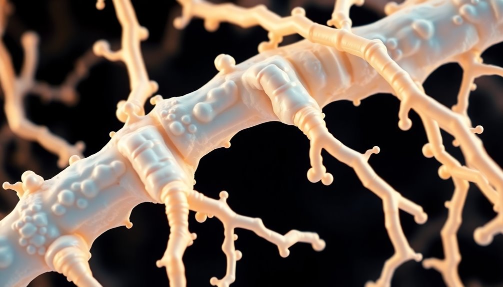
Collagen Type X is essential for bone formation, playing a significant role in regulating matrix mineralization and shaping skeletal structures. This type of collagen is vital for the development of the hypertrophic zone in bones, necessary for longitudinal bone growth. Specifically found in primary and secondary ossification centers, Collagen Type X is a key player in bone tissue development at different stages. Understanding its significance in endochondral ossification processes provides valuable insights into skeletal development mechanisms. Keep exploring to uncover more about the intricate relationship between Collagen Type X and bone formation.
Key Takeaways
- Collagen X crucial for hypertrophic zone formation in endochondral ossification.
- Essential for longitudinal bone growth and primary/secondary ossification centers.
- Plays a vital role in bone matrix mineralization during skeletal development.
- Mutations in collagen X linked to various skeletal disorders and diagnostic insights.
- Selective assembly and remodeling in bone tissues, impacting bone formation processes.
Endochondral Ossification Process
Understanding the endochondral ossification process is vital for comprehending how bones develop and grow. This intricate process involves the transformation of growth plate cartilage into bone tissue. Hypertrophic chondrocytes play a pivotal role in this process by enlarging and mineralizing the cartilage matrix components.
Collagen type X, a specific type of collagen, is essential in the formation of the hypertrophic zone within the growth plate cartilage. As hypertrophic chondrocytes undergo apoptosis, they leave cavities that are later invaded by blood vessels and osteoprogenitor cells, leading to the formation of bone trabeculae.
This process is crucial for longitudinal bone growth, as it occurs at the growth plates located at the ends of long bones. Through endochondral ossification, bones not only grow in length but also in width, contributing to the overall skeletal development and structure.
Primary and Secondary Centers of Ossification
Have you ever wondered where bone formation begins in long bones? Primary ossification kicks off in the diaphysis during embryonic development, while secondary ossification takes place in the epiphysis post-birth.
The growth plate plays a crucial role in enabling longitudinal bone growth by shifting cartilage to bone. This transformation from cartilage to bone tissue is necessary for the coordinated process of ossification.
As bones mature, the closure of growth plates signifies the end of bone lengthening, marking skeletal maturity. Understanding the distinction between primary and secondary ossification centers is essential in comprehending the intricate process of how bones develop and grow.
Collagen type X plays a significant role in this process by contributing to the structural framework required for proper bone formation at both primary and secondary ossification sites. By grasping the dynamics of these centers and their involvement in bone tissue development, we gain insight into the foundation of skeletal growth and maturation.
Fracture Healing and Bone Tissue Anatomy
Fracture healing involves a complex process known as endochondral ossification, necessary for the complete recovery of fractured bones. This intricate mechanism occurs within the fracture gap and external to the periosteum, promoting bone tissue regeneration. Histological and molecular events in long bone development are key in this healing process. Understanding the anatomy of bone tissue is vital for effectively evaluating and managing fractures.
The shoulder anatomy, specifically, is significant for studying bone tissue healing and biomechanics in the upper limb.
During fracture healing, the histological events within the bone tissue and the molecular signaling pathways that drive regeneration are essential. These processes mirror the intricate nature of long bone development and contribute to the restoration of bone strength and functionality. By delving into the details of bone tissue anatomy, healthcare professionals can gain insights into the underlying mechanisms of fracture healing and tailor treatment strategies accordingly, especially in regions like the shoulder known for their complex anatomy and functional demands.
Etymology and Publication References
Within the domain of bone formation and fracture healing, delving into the etymology and publication references surrounding endochondral ossification offers valuable insights into the process.
The term "endochondral ossification" originates from Greek, with "endo" meaning "within" and "chondral" referring to "cartilage." This term signifies the process of bone formation within cartilage, highlighting the pivotal role of cartilage in skeletal development.
Distinguished authors such as Cowan, Kahai, and Blumer have contributed significantly to the understanding of endochondral ossification, while works by Sadler, Pawlina, and Mescher provide detailed insights into this intricate process.
The etymology of the term sheds light on the complex relationship between cartilage and bone formation, emphasizing the significance of collagen type X in this biological phenomenon. These references and publications stand as foundational resources for those studying endochondral ossification, enriching the comprehension of bone development and healing processes.
Key Research Findings
Type X collagen emerges as a pivotal player in bone formation, serving as an essential marker for new bone growth and exerting significant influence on endochondral ossification. Research findings indicate that type X collagen plays an essential role in regulating matrix mineralization and organizing matrix components during bone development.
Mutations in type X collagen have been linked to conditions such as Schmid metaphyseal chondrodysplasia, affecting both the skeletal and hematopoietic systems. Type X collagen expression is specifically localized to the hypertrophic zone of the growth plate and the calcified zone of articular cartilage, emphasizing its importance in bone formation.
Additionally, elevated levels of type X collagen biomarkers are being investigated in rheumatological disorders, suggesting its potential as a diagnostic tool for bone and cartilage-related issues. These key research findings underscore the significance of type X collagen in bone formation and its implications for understanding and diagnosing various skeletal conditions.
Role of Type X Collagen
Amidst the complex process of bone formation, type X collagen stands out as an essential element produced by hypertrophic chondrocytes in the growth plate. This collagen type plays a key role in endochondral ossification, where it acts as a marker for new bone formation and regulates matrix mineralization.
By compartmentalizing matrix components, type X collagen facilitates the shift from cartilage to bone tissue, vital for proper skeletal development. The synthesis of type X collagen is closely intertwined with the process of endochondral ossification, highlighting its significance in bone formation.
Mutations in type X collagen have been linked to various skeletal disorders, underscoring the significant role this collagen plays in maintaining skeletal health. Understanding the role of type X collagen in bone formation provides valuable insights into the mechanisms governing skeletal development and the onset of skeletal disorders.
Collagen X Studies
In the domain of bone formation, studies examining collagen X have shed light on its vital role in the intricate process of endochondral ossification. Type X collagen, primarily found in the hypertrophic chondrocytes of the growth plate, is essential for bone development. Research has demonstrated that collagen X serves as a marker for new bone formation, regulating matrix mineralization and influencing the extracellular matrix composition.
Moreover, investigations have revealed how type X collagen plays a significant role in compartmentalizing matrix components, contributing to the structural integrity of the bone. By synthesizing collagen X, hypertrophic chondrocytes actively participate in the process of endochondral ossification, setting the stage for efficient bone formation.
The correlation between collagen X synthesis and endochondral ossification highlights the significance of this collagen type in orchestrating the intricate balance required for proper bone development. Through these studies, the crucial involvement of collagen X in bone formation continues to be unraveled, offering insights into potential therapeutic interventions for bone-related disorders.
Clinical Applications of Type X Collagen
The clinical applications of collagen X in bone formation showcase its promising potential for enhancing fracture healing and bone development. Type X collagen plays a pivotal role in inducing endochondral ossification, essential for effective fracture healing in synovial joints. Its involvement in the adaptive remodeling of the mandibular condyle serves as a dependable marker for new bone formation.
By regulating matrix mineralization and compartmentalizing matrix components, collagen X demonstrates clinical utility in bone development processes. The correlation between type X collagen synthesis and endochondral ossification underscores its importance in bone healing. Understanding the role of collagen X can lead to improved diagnostic strategies and therapeutic interventions for bone and cartilage disorders.
Therefore, the clinical applications of type X collagen extend beyond fracture healing to encompass its role in new bone formation, matrix mineralization, and endochondral ossification, offering potential avenues for enhancing bone development and healing processes.
Selective Assembly and Remodeling of Collagens
Among the intricate processes governing bone development, the selective assembly and remodeling of collagens stand out as essential mechanisms driving skeletal growth and structural integrity. Collagens II and IX play a key role in the chondrocyte hypertrophic phenotype, necessary for bone development. The distribution of matrical proteins in neonatal mouse mandibular condyle underscores the significance of collagens in skeletal growth.
Significantly, mice with a dominant interference mutation in collagen type X exhibit craniofacial abnormalities, highlighting its importance in bone formation. In organ culture, the differentiation of mouse embryonic mandible and squamo-mandibular joint heavily relies on collagen assembly and remodeling processes.
Collagen synthesis and remodeling are critical for maintaining the structural integrity and functionality of bone tissues. Understanding the selective assembly and remodeling of collagens, particularly collagen type X, is important for unraveling the intricate mechanisms that drive skeletal growth and contribute to preventing craniofacial abnormalities.
Conclusion
To sum up, collagen type X plays a vital role in bone formation, specifically in the process of endochondral ossification. Through its selective assembly and remodeling, collagen X contributes to the formation of the extracellular matrix in the growth plate, ultimately leading to the development of robust and healthy bones. Understanding the significance of collagen X in bone formation is crucial for advancing research and developing clinical applications in the field of orthopedics.


